Muscles
Superficial posterior compartment
More information Image, Muscle ...
| Image | Muscle | Origin | Insertion | Innervation | Main Action ! |
 |
Gastrocnemius | Lateral head: lateral aspect of lateral condyle of femur
Medial head: popliteal surface of femur; superior to medial condyle | Posterior surface of calcaneus via calcaneal tendon | Tibial nerve
(S1, S2) | Plantarflexes ankle when knee is extended; raises heel during walking; flexes leg at knee joint |
 |
Plantaris | Inferior end of lateral supracondylar line of femur; oblique popliteal ligament | Weakly assists gastrocnemius in plantarflexing ankle |
 |
Soleus | Posterior aspect of head and superior quarter of posterior surface of fibula; soleal line and middle third of medial border of tibia; and tendinous arch extending between the bony attachments | Plantarflexes ankle independent of position of knee; steadies leg on foot |
Close
[1]
Deep posterior compartment
More information Image, Muscle ...
| Image | Muscle | Origin | Insertion | Innervation | Main Action |
 |
Flexor hallucis longus muscle | Inferior two-thirds of posterior surface of fibula; inferior part of interosseous membrane | Base of distal phalanx of big toe (hallux) | Tibial nerve
(S1, S2) | Flexes big toe at all joints; weakly plantarflexes ankle; supports medial longitudinal arch of foot |
 |
Tibialis posterior muscle | Interosseous membrane; posterior surface of tibia inferior to soleal line; posterior surface of fibula | Tuberosity of navicular, cuneiform, cuboid, and sustentaculum tali of calcaneus; bases of 2nd, 3rd, and 4th metatarsals | Tibial nerve
(L4, L5) | Plantarflexes ankle; inverts foot |
 |
Flexor digitorum longus muscle | Medial part of posterior surface of tibia; by a broad tendon to fibula | Bases of distal phalanges of lateral four digits | Tibial nerve
(S1, S2) | Flexes lateral four digits; plantarflexes ankle; supports longitudinal arches of foot |
 |
Popliteus muscle | Lateral surface of lateral condyle of femur and lateral meniscus | Animation. Posterior surface of tibia, superior to soleal line | Tibial nerve
(L4, L5, S1) | Weakly flexes knee and unlocks it by rotating femur 5 deg on fixed tibia; medially rotates tibia of unplanted limb |
Close
[2]
[3]
Innervation
The posterior compartment of the leg is supplied by the tibial nerve.


 Superficial posterior compartment. Animation.
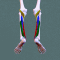 Deep posterior compartment. Animation.
